안녕하세요. 경희대 생명과학부 유전공학전공 홈페이지에 방문해주셔서 감사합니다.
현재 동문회 메뉴에 대한 업데이트를 계획 중에 있습니다.
동문회 메뉴에서 가장 주된 메뉴는 아마도 동문회 주소록이 아닐까 합니다.
부족한 실력에 어떻게든 졸업한 동문 선배님들과 재학생간의 원활한 연결을 위한 의도로 시작했지만,
검색기능에 대한 부족한 옵션과 각 동문회원의 빈약한 정보로 인해 그 기능을 다하지 못하는 것 같습니다.
동문선배님과 경희대 유전공 재학생 학우님들의 동문회 주소록 수정을 위한 조언을 구합니다.
어떤 정보가 추가 되었으면 좋겠고, 또한 검색은 어떻게 되면 간편하고 유용할 것 같은지 의견을 듣고자 합니다.
아래의 게시판에서 의견을 수렴하고 있습니다. 많은 참여 부탁드립니다. 의견수렴 게시판 가기!
View Article
Name
2005-03-03 16:51:41 | Hit : 42289 | Vote : 8340
Subject
Use of the Hemacytometer for the Determination of Cell Numbers
A hemacytometer (also spelled hemocytometer) is an etched glass chamber with raised sides that will hold a quartz coverslip exactly 0.1 mm above the chamber floor. The counting chamber is etched in a total surface area of 9 mm2 (see Figure 1).
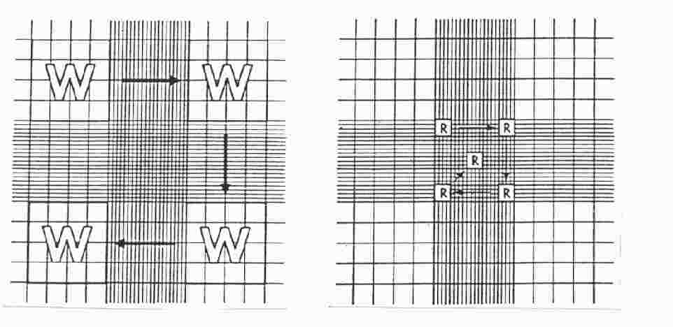
Calculation of concentration is based on the volume underneath the cover slip. One large square (see W in Figure 2) has a volume of 0.0001 ml (length x width x height; i.e., 0.1 cm x 0.1 cm x 0.01 cm).
In both methods, the hemacytometer is filled by capillary action - place the pipette filled with a well-suspended mix of cells at the notch at the edge of the hemacytometer and then slowly expel some contents so that the fluidis drawn into the chamber by capillary action.
Staining of cells often facilitates visualization and counting. Either mix cells with an equal volume of trypan blue [0.4% (w/v) tyrypan blue in PBS] to determine live/dead count (dead cells are blue) or kill cells with 10% formalin and then stain with trypan blue or other stain (to improve visualization of all cells.
Here are two simple methods for counting cells based on the surface area of the hemacytometer used to determine cell count. Other counting schemes are accetable also. The choice of methods depends upon the cell concentration - the accuracy of the procedure depends upon the number of cells counted. When cell concentration is low, one should count more grids.
Method A
Count the number of cells in the 4 outer squares (see the left panel of Figure 2).
The cell concentration is calculated as follows:
Cell concentration per milliliter = Total cell count in 4 squares x 2500 x dilution factor
Example: If one counted 450 cells after diluting an aliquot of the cell suspension 1:10, the original cell concentration = 450 x 2500 x 10 = 11,250,000/ml
Method B
Estimate cell concentration by counting 5 squares in the large middle square (see the right panel in Figure 2).
The cell concentration is calculated as follows:
Cell concentration per milliliter = Total cell count in 5 squares x 50,000 x dilution factor
Example: If one counted 45 cells after diluting an aliquot of the cell suspension 1:10, the original cell concentration = 45 x 50,000 x 10 = 22,500,000/ml
Use of the Hemacytometer for the Determination of Cell Numbers
Counting cells by the use of a hemacytometer is a convenient
and practical method of determining cell numbers in the case that the
Coulter counter is out-of-order temporarily. (It is not that bad.)
The hemacytometer consists of two chambers, each of which is divided
into nine 1.0 mm squares. A cover glass is supported 0.1 mm over
these squares so that the total volume over each square is
1.0 mm x 0.1 mm or 0.1 mm3, or 10-4 cm3. Since 1 cm3 is approximately
equivalent to 1 ml, the cell concentration per ml will be the average
count per square x 104.
Hemacytometer counts are subject to the following sources of error:
1. Unequal cell distribution in the sample
2. Improper filling of chambers (too much or too little)
3. Failure to adopt a convention for counting cells in contact with
the boundaries lines or with each other (be consistent)
4. Statistical error
With careful attention to detail, the overall error can be reduced to
about 15%. It is assumed that the total volume in the chamber represents
a random sample. This will not be a valid assumption unless the
suspension consists of individual well-separated cells.
Cell distribution in the hemacytometer chamber depends on the particle
number, not particle mass. Thus, cell clumps will distribute in the
same way as single cells and can distort the result. Unless 90% or more
of the cells are free from contact with other cells, the count should be
repeated with a new sample. A sample will not be representative if the
cells are allowed to settle before a sample is taken. Always mix the
cell suspension thoroughly before sampling.
The cell suspension should be diluted so that each such square
has between 20 - 50 cells (2-5 x 10 5 cells/ml). A total of
300 - 400 cells should be counted, since the counting error is approximated
by the square root of the total count. A common convention is to
count cells that touch the middle lines (of the triple lines) to the
left and top of the square, but do not count cells similarly located
to the right and bottom.
Hemacytometer counts do not distinguish between living and dead cells.
A number of stains are useful to make this distinction. Trypan blue
among others (Erythrosin B, Nigrosin) can be used: the nuclei of damaged
or dead cells take up the stain. If more than 20% of the nuclei are
stained, the result is probably significant. Although the trypan stain
distinction has been questioned, it is simple and gives a good approximation.
Materials
1. Clean hemacytometer and cover glass, or cover slips
2. Pasteur Pipets or Transfer Pipets
3. Balanced Salt Solution (BBS) or PBS
4. Trypan blue, 0.4% in BBS (or PBS)
5. Microscope
5. Tubes
6. Hand counter (Colony counter can be used)
7. Cell suspension
Procedure
1. Dilute 0.2 ml of Trypan blue with 0.8 ml of BBS.
2. Place cover glass over hemacytometer chamber.
3. Transfer 0.5 ml of agitated cell suspension to a 15 ml tube
and add 0.5 ml of diluted trypan blue.
4. With a Pasteur or transfer pipet, fill both chambers of the
hemacytometer (without overflow) by capillary action. Cells will
settle in the tube and in the pipet by gravity within a few seconds.
Work quickly.
5. Using the microscope with a 10X ocular (and a 10X objective),
count the cells in each of 10 squares (1 mm2 each). If over 10% of
the cells represent clumps, repeat entire sequence. If fewer than
200 or more than 500 cells are present in the 10 squares, repeat with
a more suitable dilution factor.
6. Calculate the number of cells per ml, and the total number of
cells, in the original culture as follows:
Cells/ml = average count per square x 104
Total cells = cells per ml X any dilution factor X total volume of
cell preparation from which the sample was taken.
7. Repeat count to check reproducibility (+/- 15%).
References
1. Berkson, J., T. B. Magath and M. Hurn (1939). Am. J. Physiol. 128, 309.
2. Sanford. K.K., W.R. Earle, V.J. Evans, H.K. Waltz and J.E. Shannon (1951).
3. Absher, M. in Tissue Culture Methods and Applications, Eds. Kruse, P.F. and Patterson, M.K., Jr. Academic Press, N.Y., 1973, p.395.
From the Laboratory of Dr. Allan Bradley
Baylor College of Medicine, Houston, Texas
Copyright 1998 (c) 경희대학교 생명과학대학 생명과학부 유전공학전공.
(우) 449-701 경기도 용인시 기흥읍 서천리 1번지. Tel : 031 - 201 - 2433 | Fax : 031 - 203 - 4969
All rights reserved. 홈페이지 최종수정일 : 2005년 1월 3일 03:52
- Counting status :
visited today,
visited yesterday,
visited in sum.


 visited today,
 visited yesterday,
 visited in sum.